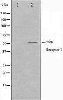
![Western blot analysis of TNFR1 Antibody expression in A549 cells lysates.The lane on the left is treated with the antigen-specific peptide.](http://www.phscientific.com/cdn/shop/products/909_thumb_P_1406632814747_{width}x.jpg?v=1591794989)
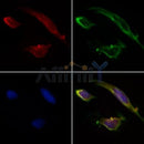
![AF0282 staining HepG2 by IF/ICC. The samples were fixed with PFA and permeabilized in 0.1% Triton X-100,then blocked in 10% serum for 45 minutes at 25¡ãC. Samples were then incubated with primary Ab(AF0282 1:200) and mouse anti-beta tubulin Ab(T0023 1:200) for 1 hour at 37¡ãC. An AlexaFluor594 conjugated goat anti-rabbit IgG(H+L) Ab(S0006 1:200 Red) and an AlexaFluor488 conjugated goat anti-mouse IgG(H+L) Ab(S0017 1:600 Green) were used as the secondary antibod](http://www.phscientific.com/cdn/shop/products/af0282_tnfr1_antibody_thumb_P_1547375623473_{width}x.jpg?v=1591794989)
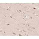
![AF0282 at 1/100 staining human brain tissue sections by IHC-P. The tissue was formaldehyde fixed and a heat mediated antigen retrieval step in citrate buffer was performed. The tissue was then blocked and incubated with the antibody for 1.5 hours at 22¡ãC. An HRP conjugated goat anti-rabbit antibody was used as the secondary](http://www.phscientific.com/cdn/shop/products/909_thumb_P_1512439188740_{width}x.jpg?v=1591794989)
![Western blot analysis of TNFR1 Antibody expression in A549 cells lysates.The lane on the left is treated with the antigen-specific peptide.](http://www.phscientific.com/cdn/shop/products/909_thumb_P_1406632814747_130x.jpg?v=1591794989)
![AF0282 staining HepG2 by IF/ICC. The samples were fixed with PFA and permeabilized in 0.1% Triton X-100,then blocked in 10% serum for 45 minutes at 25¡ãC. Samples were then incubated with primary Ab(AF0282 1:200) and mouse anti-beta tubulin Ab(T0023 1:200) for 1 hour at 37¡ãC. An AlexaFluor594 conjugated goat anti-rabbit IgG(H+L) Ab(S0006 1:200 Red) and an AlexaFluor488 conjugated goat anti-mouse IgG(H+L) Ab(S0017 1:600 Green) were used as the secondary antibod](http://www.phscientific.com/cdn/shop/products/af0282_tnfr1_antibody_thumb_P_1547375623473_130x.jpg?v=1591794989)
![AF0282 at 1/100 staining human brain tissue sections by IHC-P. The tissue was formaldehyde fixed and a heat mediated antigen retrieval step in citrate buffer was performed. The tissue was then blocked and incubated with the antibody for 1.5 hours at 22¡ãC. An HRP conjugated goat anti-rabbit antibody was used as the secondary](http://www.phscientific.com/cdn/shop/products/909_thumb_P_1512439188740_130x.jpg?v=1591794989)
Description
Delivery Information
Payment & Security
Your payment information is processed securely. We do not store credit card details nor have access to your credit card information.